Issue 14, August 22, 2016
Bur Oak Blight and Look-alike Diseases
The U of I Plant Clinic has received many samples and questions regarding diseased bur oak trees. Most clients were concerned about possible Bur Oak Blight or Oak Wilt infections. This article will address some of the key diagnostic symptoms for Bur Oak Blight and some of its look-alikes.
Bur Oak Blight
Bur Oak Blight (BOB) is a fungal disease caused by the pathogen Tubakia iowensis. An important characteristic of BOB is that it is a late season leaf blight. The most notable symptoms first appear in late July and August. Leaf blight symptoms appearing earlier than that are not likely to be related to BOB. Initially, infections are limited to foliage in the lower canopy. Infections progressively expand upwards, throughout the canopy from year to year as disease inoculum (fungal spores) within the tree builds. When scouting for BOB look for diseased leaves with a characteristic wedge shaped necrotic lesion near the apex of the leaf blade (Photo 1). Coalescing lesions and expanding vein necrosis causes the leaves to die and results in extensive defoliation. A more important phase of the disease, especially in terms of the pathogen's ability to overwinter, involves an early season infection of expanding shoots, followed by necrosis of the petioles several months later, usually in late July. Leaves killed by this phase of the disease remain attached to the tree into the winter, well after healthy bur oaks have dropped their leaves. Diseased leaf retention is currently one of the best ways to identify suspect trees. The leaf blade eventually breaks off, but the petiole remains attached. Look for petioles attached from the previous growing season. Infected petioles will have black pustules or scars from previously attached pustules (Photo 2). The diseased petioles produce the spores responsible for primary infections. The U of I Plant Clinic looks for combination of three symptoms and signs when diagnosing BOB: 1) wedge-shaped necrosis on the leaf blade, 2) Tubakia spp. on the leaf blade (microscope needed), and 3) a branch with diseased petioles still attached from the previous growing season. Refer to Issue 16, September 16, 2015 for more information on Bur Oak Blight.
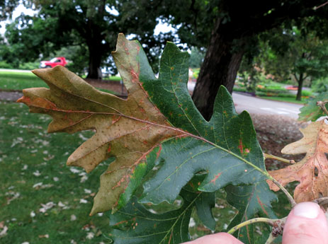
Photo 1. Wedge-shape lesion characteristic of Bur Oak Blight
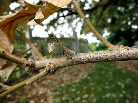
Photo 2. Branch with diseased petiole still attached from previous growing season. Note the small black pustules.
Tubakia Leaf Spot
Tubakia Leaf Spot is a common late season (July and August) leaf disease caused by the pathogen Tubakia dryina, which is closely related to that of Bur Oak Blight. The disease is more common on members of the red oak group (Photo 3), but does cause frequent infections to bur oak. Unlike Bur Oak Blight, Tubakia Leaf Spot is considered a minor disease. Tubakia leaf spot lesions will vary with host susceptibility and environmental conditions. The lesions start as small water soaked areas. They become evident as they enlarge and transition to a reddish brown color. Severe infections can cause premature leaf drop. The Tubakia pathogen is fairly easy to confirm in a diagnostic laboratory with the aid of a microscope. Tubakia Leaf Spot is not associated with tree mortality and is not known to infect or produce overwintering pustules on petiole tissues. Refer to Issue 13, August 3 2015 for more information on Tubakia Leaf Spot.
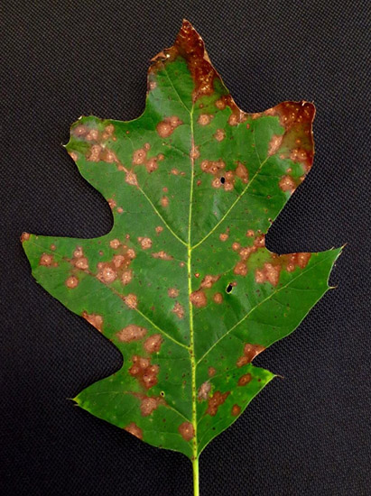
Photo 3. Northern red oak leaf with numerous Tubakia Leaf Spot lesions.
Oak Wilt
Oak Wilt was covered in Issue 12 of this year's newsletter. As a review, bur oak, and other members of the white oak group, are less susceptible to Oak Wilt. In fact, the plant clinic rarely confirms oak wilt on members of the white oak group. Infections on this group typically are slow to progress, and are confined to scattered branches of the canopy. This may help differentiate from Bur Oak Blight, which is initially concentrated on the lower canopy. Leaves on oak wilt infected branches appear wilted and have a light brown or straw-colored discoloration that moves inward from the leaf margin. While infected leaves may remain attached to the branches, they lack the leaf vein and petiole symptoms seen with Tubakia spp. (Travis Cleveland)
Author:
Travis Cleveland