Issue 10, June 25, 2012
Have You Ever Wondered How the U of I Plant Clinic Cultures and Isolates for Vascular Pathogens?
When someone suspects that their tree is dying from a vascular pathogen such as Oak wilt, Dutch elm disease, or Verticillium wilt, they can send a sample to the U of I Plant Clinic. We usually suggest that they sample from areas of the trees that are showing early symptoms typical of disease as well as wood that may show streaking or darkening of vascular tissue (see pictures below). We would like the sample to consist of several 1 to 2 foot long branches with at least the diameter of a thumb. Branches with diameters smaller than ½" are difficult to culture and may not provide accurate results. Contact the U of I Plant Clinic for instructions on how to correctly collect and submit your samples. Our contact information can be found on the Plant Clinic website.
The procedures of culturing various vascular pathogens are very similar. Below is an example of how we culture for Oak wilt in our plant diagnostic lab.
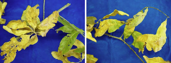
Some oak leaves showing symptoms of oak wilt.
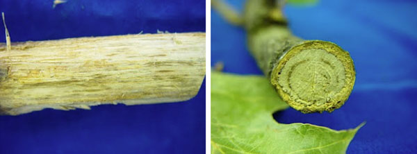
Dark streaking of the wood caused by the oak wilt fungal pathogen.
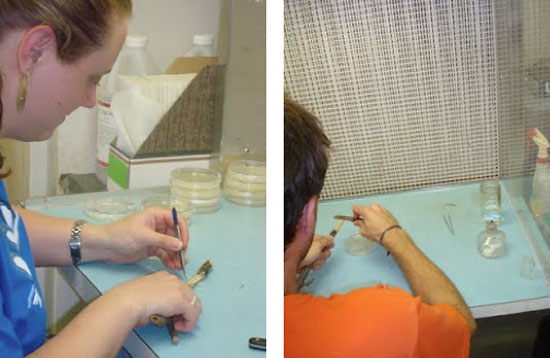
Here students at the U of I Plant Clinic:
- Take notes on the condition of the oak sample.
- Label agar plates with sample number, date, type of agar, and OW (Oak Wilt)
- The bark is peeled back from the end of the branch, so that the wood is exposed under a sterile hood.
- They flame their knife and notch the wood into tiny wood chips, which remain attached to the branch.
- Then, they flame their tweezers and pick off wood chips.
- These wood chips are placed, very quickly, (to avoid any unwanted contamination) into PDA -Potato Dextrose Agar, plates under a sterile hood.
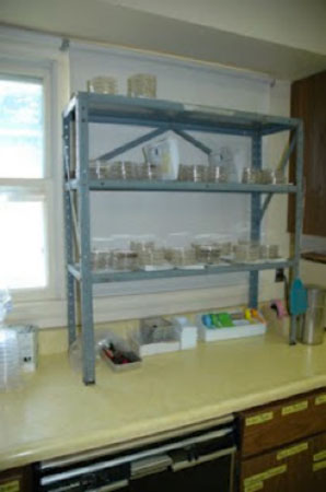
The agar plates are stored on a shelf in our lab, with their paperwork. They are kept there for up to 7 to 14 days to allow time for the fungal isolates to grow.
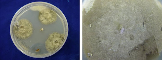
Pictures of the oak wilt fungus growing on a culture plant with PDA
If we were successful at isolation of the oak wilt fungus, the above pictures show what it would look like growing in our PDA culture plates.
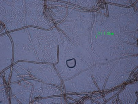
Picture of oak wilt spores in chains taken by Travis Cleveland
Lastly, we take clear tape and place it on top of the fungus to "catch spores". This clear tape is placed onto a microscope slide with a drop of water on it. If the oak tree is infected with oak wilt or positive for oak wilt, we will see the chains of spores that are seen in the picture above. If none of these spores can be found, the oak sample will be considered "oak wilt negative." (Stephanie Porter and Travis Cleveland)
Authors:
Stephanie Porter
Travis Cleveland